Foot and ankle pain is a prevalent problem that affects many people’s ability to enjoy daily activities, exercise and play sports. Justin Price covers the basic anatomy of the feet and ankles, explains the most common musculoskeletal imbalances in these areas, teaches you how to assess the feet and ankles, and provides two corrective exercises for foot and ankle pain to help sufferers feel and function better.
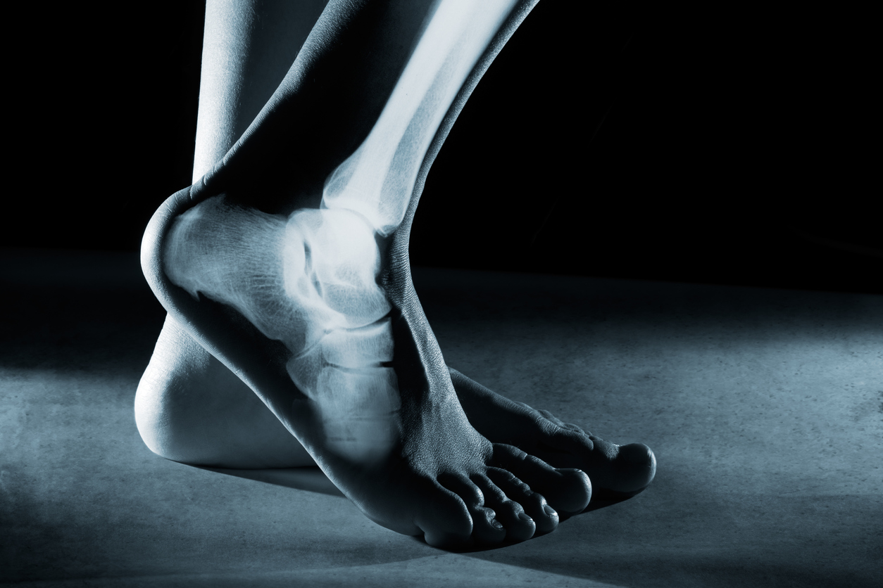
The feet and ankles are key parts of the body that act as shock absorbers when you interact with a contact surface such as the ground. They also help the body adapt to varied surfaces. Understanding the anatomy of these important body parts helps you to know how to assess them for problematic imbalances.
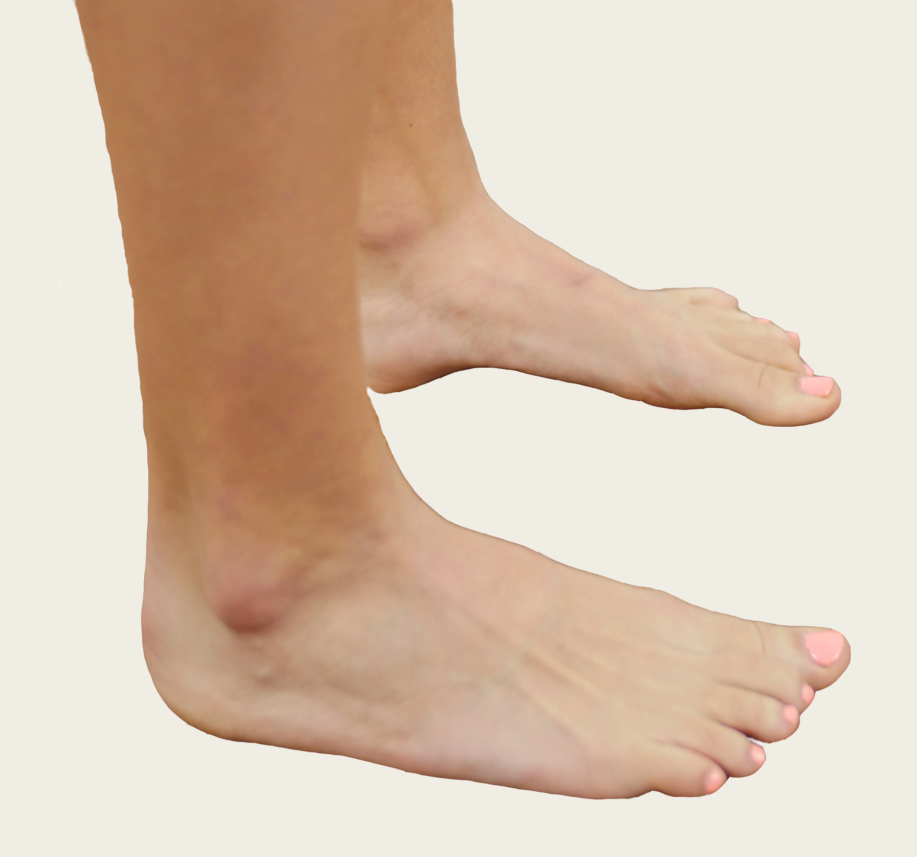
The ankle can be divided into two parts: the true ankle joint and the subtalar joint. The true ankle joint is immediately below the shin bones and is responsible for up and down movement of the foot. The subtalar joint is located below the true ankle joint, between the talus bone and the heel bone.1,2 This joint helps transfer weight from side to side (see Figure 1).
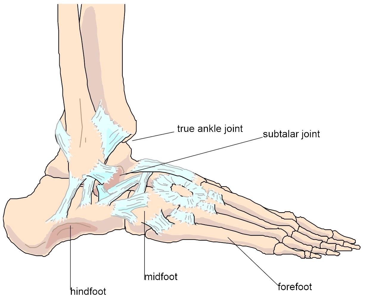
Figure 1: Anatomy of the foot and ankle
The foot consists of three parts: the hindfoot, the midfoot and the forefoot. The hindfoot (talus and heel bone) acts to absorb shock and displace it forward and from side to side. The midfoot (small bones in the foot between the heel and the toes) helps continue dissipating force, while the forefoot (toes) adapts further to the terrain and aids propulsion during gait (see Figure 1).1,2
Common imbalances that can cause foot and ankle pain
The most common imbalances found in the feet and ankles that can cause pain are:
- Overpronation
- Lack of dorsiflexion
Pronation is a necessary function of the foot. It is needed to help transfer weight forwards and towards the midline of the body. Overpronation is when the foot collapses too much. This can affect the correct function of the feet, ankles and rest of the body.
Dorsiflexion happens when the foot moves towards the shin and vice versa. The ability to dorsiflex can be impacted as a result of overpronation. Overpronation causes the foot, ankle (and knee) to roll inward too much, which limits the ability of the shin to travel forwards over the foot (i.e., dorsiflex). Limited dorsiflexion impairs all weight-bearing activities, from standing to squatting to walking and running.
Foot and ankle assessment
You must be able to see and feel the feet, ankles and knees clearly in order to assess these structures properly. Ask clients to remove their shoes and socks for this assessment.
This hands-on assessment of your client’s subtalar joint will help you evaluate for overpronation. Locate the two small indentations at the base of your client’s ankle, just below the prominent ankle joint. Place your thumb on the dimple on the inside of the ankle and your forefinger on the dimple on the outside (see Figure 2).4 Ask your client to roll their foot and ankle inward (overpronate). You will feel pressure increase on your thumb. Ask them to roll outward (oversupinate) and you will feel pressure increase on your forefinger. This pressure is the talus bone moving in the ankle. Coach your client to pronate and supinate until you feel an even amount of pressure of the talus bone on both your thumb and forefinger. This is the anatomical neutral position for the foot and ankle when standing.5
Note: Most people will have to supinate to get to neutral from their dysfunctional overpronated position.
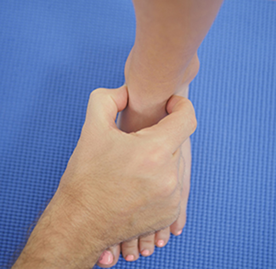
Figure 2: Assessing the foot and ankle
The relationship between the feet and ankles and the knees
While your client is in a neutral foot and ankle position, look at their knees. The centre of the kneecap will now likely be in line with the second toe (its anatomical neutral position). When your client relaxes from neutral, observe the movement of their knees as they fall back into their more comfortable (likely overpronated) foot position. Notice how their knees rotate inward with their foot, ankle and shins. This rotation can lead to problems in their feet and ankles, as well as their knees.3
Corrective exercises for foot and ankle pain
Squats, lunges, running and walking all involve motion of the foot and ankle. Musculoskeletal imbalances in these areas, such as overpronation and a lack of dorsiflexion, can lead to foot, ankle and leg problems like plantar fasciitis, Achilles tendinitis, bunions and shin splints. Here are two exercises to help your clients correct these structural deviations.
Golf Ball Roll
Overpronation leads to wear and tear of the plantar fascia and degeneration of structures on the underside of the foot. The Golf Ball Roll is a myofascial massage technique which can help your clients regenerate the tissue on the underside of their feet.
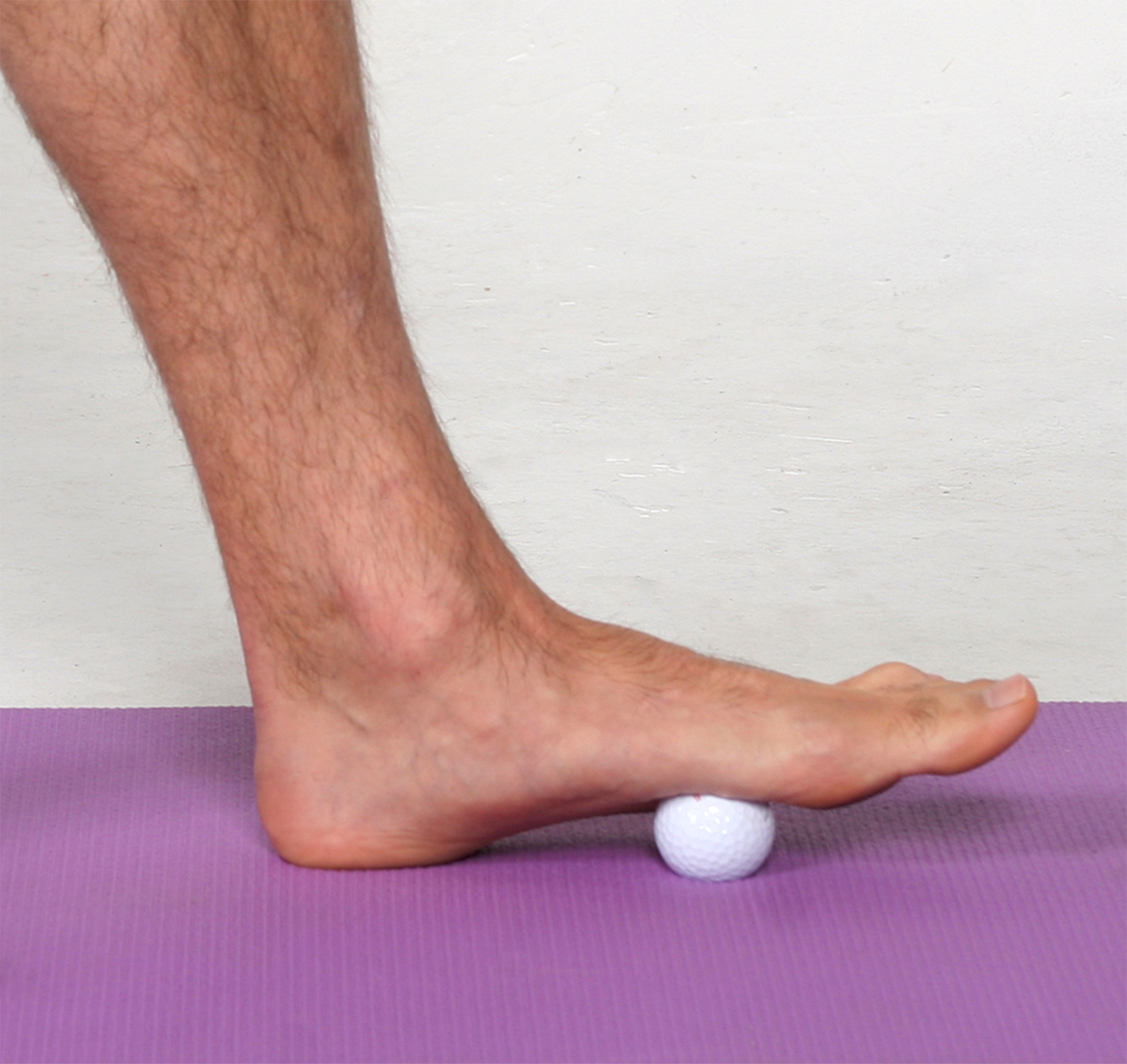
Golf Ball Roll
Roll a golf ball daily on the underside of each foot for 30 seconds to one minute on the sore spots they find at least once a day.
Big Toe Pushdowns
When people overpronate, the arch of their foot becomes weak. The Big Toe Pushdown exercise helps strengthen the arch so that it can help support the foot correctly.
Big Toe Pushdowns
Instruct your client to find a neutral foot and ankle position (see Figure 2: Technique for assessing foot and ankle position). Ask them to maintain this position as they push their big toe down without collapsing the arch of their foot. As they get stronger, they will feel the muscles of the arch contract under their foot. You can coach your client to activate this muscle in all weight-bearing exercises to prevent overpronation.
Assessments and corrective exercises for foot and ankle pain can be integrated into any fitness programme. Simply incorporate exercise strategies that address any musculoskeletal imbalances you identify during the assessment process to help your clients feel better and function more effectively.
To learn more from Justin Price about how to help both yourself and your clients feel and function better, check out The BioMechanics Method Corrective Exercise Specialist course available through FitPro.
Explore our sports massage therapist insurance and get covered today.
References
- Golding LA & Golding SM (2003), Fitness Professional’s Guide to Musculoskeletal Anatomy and Human Movement, Monterey, CA: Healthy Learning.
- Gray H (1995), Gray’s anatomy, New York: Barnes & Noble Books.
- Mccreary E, Romani W, Rodgers M, Kendall F & Provance P (2005), Muscles Testing and Function with Posture and Pain (5th ed.), Baltimore, Md: Lippincott Williams and Wilkins.
- Price J & Bratcher M (2019), The BioMechanics Method Corrective Exercise Specialist Certification Program (2ndEdition), San Diego: The BioMechanics Press.
- Price J (2018), The BioMechanics Method for Corrective Exercise, Champaign, IL: Human Kinetics.